Retinal Photography
|
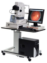
Retinal photography is a must in optometric practices. It is essential in managing glaucoma.
Photographic documentation of diabetic retinopathy patients helps us keep a data base of the progression of the disease, it's on going management and control. High Resolution Retinal Photography uses a computer-integrated digital imaging system to record a detailed view of the retina. Since nothing touches the eye, photo-documentation is painless. This digital image provides an excellent reference point for future comparisons. If you need this digital image in the future will gladly e-mail you a copy image |
|
Retinal photography assists in the detection and management of problems such as diabetic changes, hypertensive (high blood pressure) retinopathy, macular degeneration, optic nerve disease, and retinal holes, thinning and/or detachments.
We recommend that all our patients receive this test. It is especially important for people with a history of high blood pressure, diabetes, retinal diseases, flashing lights, floaters, headaches or a strong/high eyeglasses prescription.
We recommend that all our patients receive this test. It is especially important for people with a history of high blood pressure, diabetes, retinal diseases, flashing lights, floaters, headaches or a strong/high eyeglasses prescription.
Optical Coherence Tomography
|
Optical coherence tomography (OCT) is a non-invasive imaging test that uses light waves to take cross-section pictures of your retina.
With OCT, the doctor can see each of the retina’s distinctive layers. These measurements help with diagnosis and guide treatment for glaucoma as well as retinal disease, like age-related macular degeneration (AMD) and diabetic eye disease. OCT is often used to evaluate disorders of the optic nerve and allows the doctor to monitor changes to the fibers of the optic nerve to detect changes that may be caused by glaucoma. |

|
Visual Field Test
|
Visual Field Testing monitors peripheral vision. Visual fields are obtained to monitor visual changes that may be caused by specific eye diseases, such as glaucoma, as well as the neurological function of the retina, optic nerve and brain. While there are several types of visual field exams, the most common required patients to focus on one spot and respond to flashing lights by pressing a button.
Your intraocular eye pressure (IOP) is important in determining your risk for glaucoma. If you have high IOP, careful management of your eye pressure with medications can help prevent vision loss. Ours Ollyeyes VisuALL Virtual Reality Visual Field Testing is an innovative portable device that leverages the power of virtual reality and artificial intelligence to enable eye-care providers to monitor common eye diseases. |

|
Corneal Topography
|
Corneal Topography provides a mapping of your cornea. This may be required for certain eye conditions, prior to refractive surgery, or for specialty contact lenses. A corneal topographer is a computer linked to a lighted bowl with a pattern of concentric rings inside it. The patient sits at the bowl with their forehead braced against a bar. The technician has only to line the patient up properly and snap an image.
The procedure is painless and very fast. The computer then uses the snapped image to produce a printout of the corneal shape using colors to identify different depths much like a topographic map of the earth describes changes in the land surface. |

|